Innovative Nodes
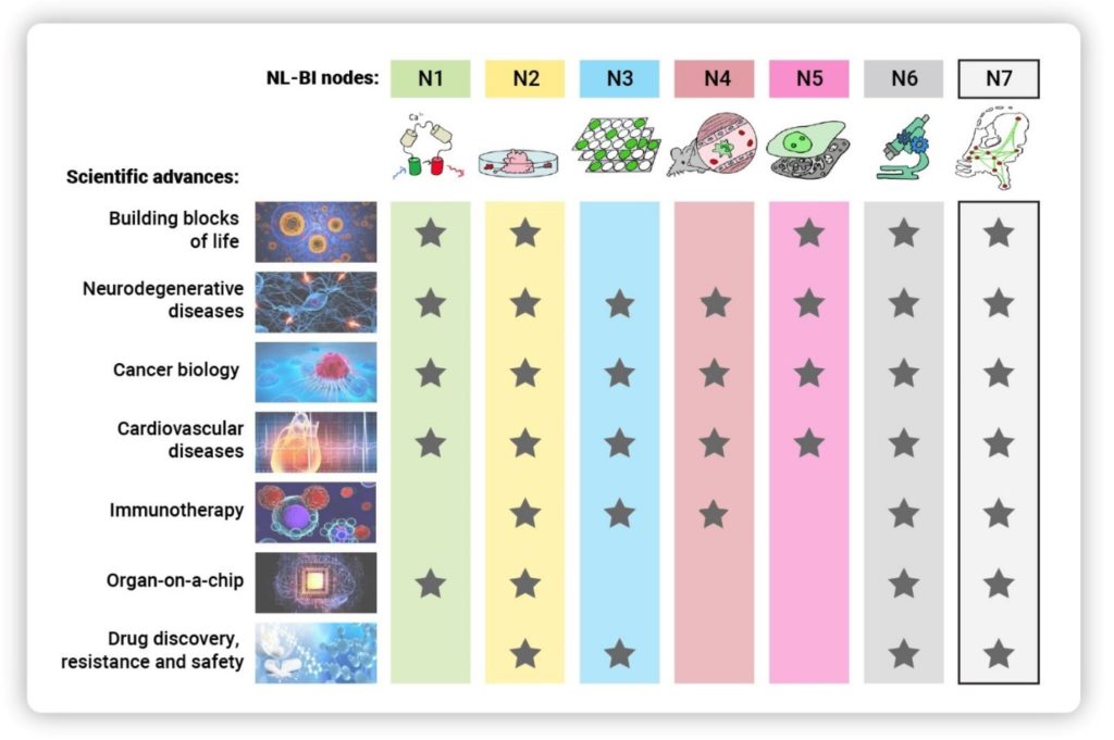
Node 1 (FP): Gadella (UvA), Giepmans (UMCG),
Houtsmuller (EMC), Jalink (NKI), Kapitein (UU)
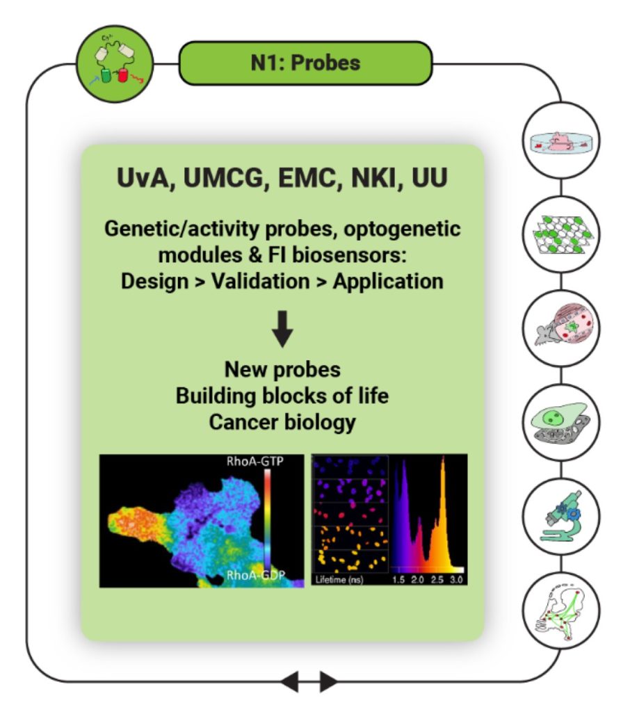
Expanding the detection toolbox by introducing novel fluorophores, molecular reporters for intensity-based and FRET/FLIM detection as well as non-linear detection of tissue-intrinsic signals using higher harmonic imaging and Raman scattering. Genetically encoded biosensors and actuators will be generated, validated, and applied to directly spy on or alter physiological changes at high resolution in real time, enabling imaging of second messenger enzyme activity in cultured cells, organoids and disease models.
Node 2 (3D- FI): Borst (WUR), Reits (A-UMC), Rios (PMC)
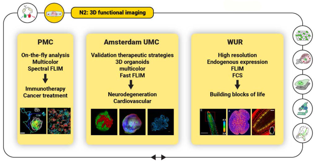
3D imaging of multicellular organisms like organoids or plants allow to obtain the landscape of molecular events and to reveal the mode of action of proteins in their natural habitat. Through multi-parameter detection, high content multi-parameter live-cell microscopy (ten or more channels) will combine fast multicolor imaging, on-the-fly data analysis and functional imaging techniques including FLIM and FCS to integrate the study of morphological, molecular and functional aspects of live-cell biology in 3D model systems.
Node 3 (HTM): Beiersbergen and Jalink (NKI), Le Dévédec (Leiden)
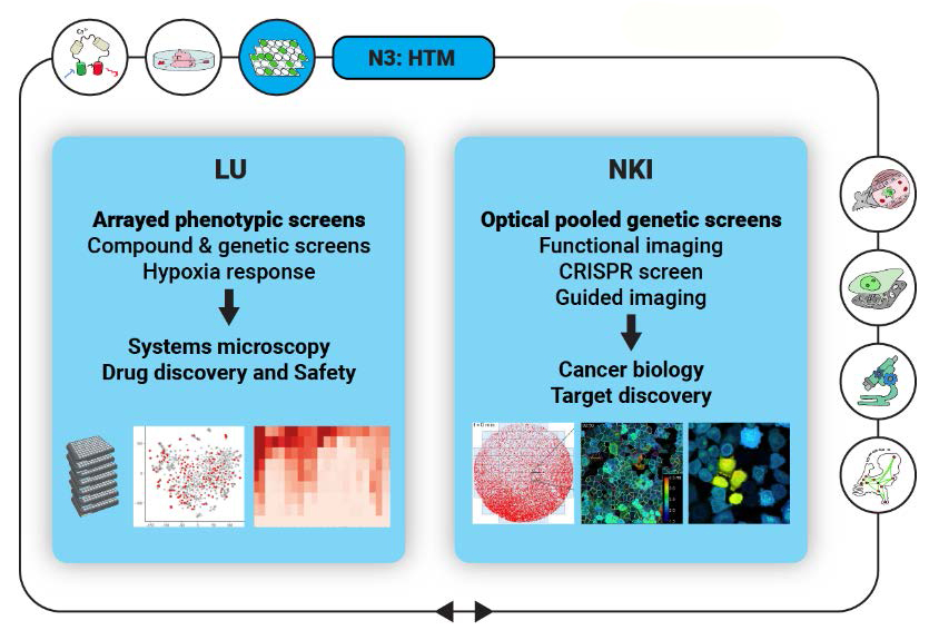
Microscopy phenotypic screens allow to uncover molecular mechanisms of disease progression, candidate drug targets, mechanisms of drug resistance, novel drug (combinations) for effective treatment, and safe therapeutic windows. By upscaling the automation in the high-throughput microscopy pipeline, users will have access to a completely automated screening pipeline from libraries aliquoting, automated imaging and image analysis. Pooled microscopy screening enables effective screening for literally any phenotype that can be detected by microscopy, be it rare morphological phenotypes, changes in dynamic parameters or even any of the functional imaging assays. Pooled microscopy screens rely heavily on microscope automation and on-the-fly image analysis and will reduce screening time and costs.
Node 4 (IVM): Friedl (RadboudUMC), van Rheenen (NKI),
Groot (VU), van Zandvoort (UM)
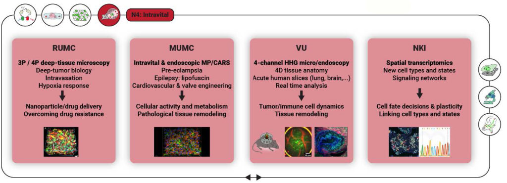
Confocal microscopy and endoscopy, light sheet, and multiphoton microscopy all allow to dynamically and in 3D examine live cells and tissue context inside small vertebrates as well as in human tissue (in situ or acutely after isolation). The obtained multi-parametric datasets on in vivo cell biology deliver precise functional information on cell type, migration activity, cell-cell interaction and alterations, including inflammatory states and tissue degeneration. This is essential in understanding pathogenesis in diseases as oncology, atherosclerosis, and neurodegeneration. Increasing imaging depth in light-absorbing, scattering tissue, using novel low repetition rate laser systems for far-red (1300-1700 nm) excitation with high-pulse energy; deep, otherwise hidden tissues can further be accessed by fiber-based and bench top endo-microscopes, using linear (multiphoton) and nonlinear (SHG, THG, CARS) excitation schemes; Imaging in a broad type of acute human tissues, introducing 3D pathology into medical research. The ultimate goal is the introduction of diagnostic microscopy into the clinical arena, thereby revolutionizing on-the-spot diagnosis and intervention in the surgery suite using fiber-based multiphoton endoscopy.
Node 5 (CLEM): Giepmans (UMCG), Hoogenboom (TUD), Klumperman and Liv (UMCU)
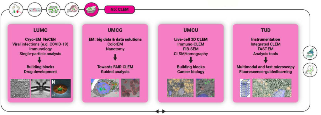
live-cell CLEM provides an exciting new tool to study cellular dynamics and life functional and molecular information in healthy and diseased conditions all the way from single proteins and organelles to EM ultrastructure. Combined with 3D models, such as organoids and whole tissues, this provides a unique and robust platform to study fundamental biological processes within their relevant context and link patient-specific genetic mutations to phenotypic information. By connecting live-cell fluorescence and intrinsic signal, such as (cryo-)CARS, microscopy with electron microscopy, 3D correlative cryo-electron microscopy will allow to identify ultrastructural changes underlying molecular mechanisms of disease
Node 6 (Smart microscopy): Houtsmuller (EMC), Jalink (NKI), Kapitein (UU), Rieger (TUD)
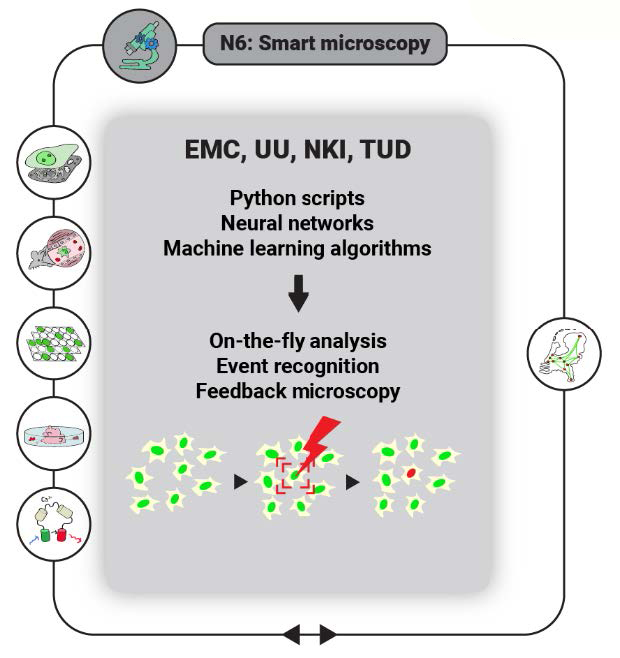
Merely visualizing cellular processes with a microscope is seldomly sufficient to fully understand them. Therefore, many quantitative and qualitative assays including the various functional imaging assays have been developed to study the intracellular behaviour of biomolecules and their interactions. In feedback microscopy, images are analysed for rare events/features on the fly, and the results are fed back to the microscopy hardware to alter the course of image acquisition or to trigger specific actions such as photoactivation of probes, induction of DNA damage by UV irradiation, or killing of rare lymphoma cells amongst many healthy blood cells. On-the-fly phenotype analysis will be used to detect rare events (such as a T cell killing a target cell), triggering adaptations to the imaging protocols, FI protocols (FRAP, FLIM), activation of external stimuli etc. This development drastically increases throughput by allowing unsupervised imaging and impacts functional imaging (FLIM, FCS, optogenetics), high-content screening (pooled microscopy screens) and intravital microscopy
Node 7 (DMDA) Data Management & Analysis: Giepmans (UMCG), Wolstencroft (LU)
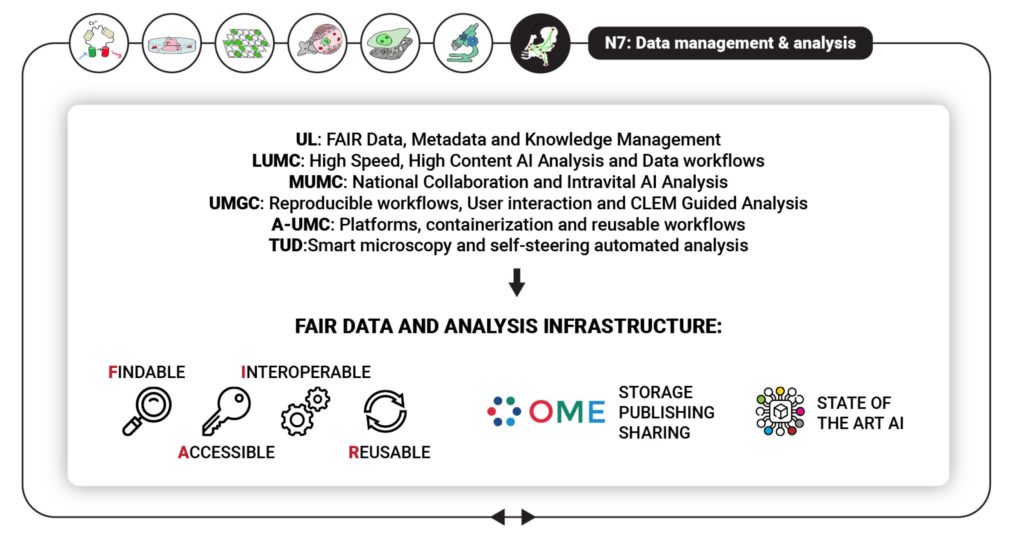
FAIR (Findable Accessible, Interoperable and Reusable) data management is a challenge for the optical microscopy communitydue to a high diversity in participating research fields and methodologies, diversity in data formats, and the large volumes of data generated. Introducing FAIR data management for reproducible, reusable results by developing a nationwide network for the FAIR sharing of data and image analysis methods, and facilitate a greater exploitation of valuable research results, increased collaboration within the Netherlands and worldwide, and enable the adoption of cutting-edge AI analysis methods for Dutch microscopy.
Node 8 Outreach and Training: Giepmans (UMCG), Reits (A-UMC) NVvM
NL-BI will enable breakthroughs by optimal usage of requested and available microscopes that are part of the NL-BI infrastructure as well as the (local) expertise from involved operators and embedded researchers. To enhance nationwide awareness and strengthen embedding and application of the newest developments in all research labs in the Netherlands, we will develop focused technology training workshops for (potential) users. This will be done via interactions and exchange within the specific NVvM working groups and by shadowing exchange of technicians amongst Core Facilities (“train the trainer”). Biannual core facility operator days will focus on specialized topics such as quality control, sample preparation, and data analysis.